|
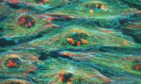
Image: Not only did researchers at UCSD and UCI visualize a mechanically induced signal traverse human cells, but they also showed that actin filaments and microtubules are involved in the process. Human cells filmed instantly messaging for first time Cells tugged in one direction sent biochemical signals in the opposite direction in the form of a signature pattern of fluorescent light Researchers at UCSD and UC Irvine have captured on video for the first time chemical signals that traverse human cells in response to tiny mechanical jabs, like waves spreading from pebbles tossed into a pond. The scientists released the videos and technical details that explain how the visualization effect was created as part of a paper published in the April 21 issue of Nature. The researchers working at the UCSD Jacobs School of Engineering's Department of Bioengineering developed a novel molecular "reporter" system, which allowed the dynamic visualization of the activation of an important protein called Src. Peter Yingxiao Wang, lead author of the paper and a post-doctoral researcher in UCSD's Jacobs School of Engineering spent two years designing the reporter molecules to light up selectively only when Src was activated, and not other proteins. Wang and his co-workers first demonstrated that the novel system was effective in visualizing Src activation in response to a known chemical stimulant, epidermal growth factor. Next, they studied the effect of mechanical stimuli on Src activation. Using technology developed at the Beckman Laser Institute at UC Irvine by its founding director Michael Berns, Wang and Elliott Botvinick, a postdoctoral researcher at UCSD Department of Bioengineering and the Beckman Laser Institute at UC Irvine, attached small, sticky beads to cells and gently tugged the beads to and fro with laser power acting as invisible "tweezers." As the laser tweezers moved the beads in one direction, a video camera attached to a specially equipped microscope recorded the dynamic movement of biochemical signals in the opposite direction in the form of a signature pattern of fluorescent light. The fine spatial and temporal resolution was made possible by a technology called fluorescence resonance energy transfer. "We had no idea what to expect," said Wang. "The first time we saw these incredible waves spreading across the cells I just said 'Whoa, this is amazing.' We expected to see a signal where the tweezers were pulling the beads, but we did not envision such a directional wave propagating away from the beads." Wang worked on this project under the joint advisorship of Shu Chien, a professor of bioengineering and medicine and director of the Whitaker Institute of Biomedical Engineering at UCSD, and Roger Y. Tsien, professor of pharmacology, chemistry, and biochemistry and investigator with the Howard Hughes Medical Institute at UCSD. Src is one of a large group of enzymes called kinases that attach a phosphate molecule to one or more target proteins in the cell. This phosphorylation reaction typically switches the target protein from inactive to active status. Many diseases can result either when a kinase gene is mutated and can't properly phosphorylate its targets, or when a normal kinase becomes overactive or not sufficiently active. Indeed, Src has been shown to play a key role in cell growth and development, and in the genesis of cancer, atherosclerosis, and many other disease conditions. "This study amounts to a proof of principle that if we can visualize the activation of one kinase, we can do the same for many others using the same approach," said Chien, the senior author of the paper. Not only are those additional studies expected to reveal temporal and spatial patterns of kinase activation, but Chien also predicted that there will be practical spin offs. For example, cells usually tightly control the activity of Src, but in certain cancers its activity is abnormality high. "We think that our ability to measure Src activity with this new visualization technique would be useful as a diagnostic test for many cancers," said Chien. The William J. von Liebig Center for Entrepreneurism and Technology Advancement at UCSD's Jacobs School has provided Chien and Wang with funding to commercialize the new visualization technology as a cancer-detection tool. The researchers showed that actin filaments and microtubules, structural elements that traverse cells like the ribs of an umbrella, could function as conduits for the spread of biochemical signals. Indeed, when Wang disrupted either actin filaments or microtubules in his test cells, the activation signal no longer spread across the cell. These results suggest that the activation of Src traverses these filamentous structures. Source: University of California - San Diego 美国加州大学的圣地亚哥分校(UCSD)及欧文分校的研究人员首次视频捕获到响应微小机械尖刺穿过人类细胞的化学信号,它们就像鹅卵石投入池塘中激起的波纹。他们的研究论文发表在2005年4月21日的《自然》杂志上,论文主要作者是UCSD雅各布斯工程学院的博士后研究员Peter Wang。 UCSD雅各布斯工程学院生物工程系的研人员开发了一种新的分子“报道”系统,这种系统能动态显示Src蛋白质激活情况。Wang花了两年时间使报道分子能够仅在Src而非其他蛋白质被激活时才发出指示。 Wang及其同事首先证明新系统能有效显示响应一种已知的化学刺激物――表皮生长因子的Src激活的情况。然后,他们研究了机械刺激对Src激活的效果。采用加州大学欧文分校贝克曼激光研究所研制出来的技术,Wang和另一位博士后研究员Elliott Botvinick将一些粘性小珠, 固定在细胞上,然后利用激光源(无形的“镊子”)来回拉动这些小珠。当激光镊子使这些小珠朝一个方向移动时,固定在一个专用显微镜上的摄像头就会拍摄下生化信号朝着相反方向的动态移动,其移动方式具有荧光的特征。因此,采用荧光共振能量转移(FRET)技术,就有可能实现高空间分辩率和高瞬时清晰度。 Wang说,当他们第一次看见这些不可思议的波纹穿越细胞扩散开来时,真是觉得太奇妙了,他们根本没想到会看见从小珠发散出来的定向波纹。 Src是一种激酶,它将磷酸盐分子固定到细胞内的一个或多个目标蛋白质上(称为“磷酸化”),从而激活目标蛋白质。如果激酶基因变异或不能正常磷酸化其目标,或激酶过于活跃,或激酶不够活跃时,都会导致多种疾病。事实上,Src在细胞生长发育,以及癌症、动脉硬化及其他许多疾病的产生中都起到重要作用。 论文另一作者Chien说,这一衡量Src活性的可视化技术可用于许多癌症的诊断。细胞通常严格控制Src的活性,但是,在某些癌症情况下,Src的活性异常高。Chien和Wang已获得将这一可视化新技术作为癌症诊断工具来商业化的基金。 这些研究人员还发现,肌纤蛋白丝和微管(横贯细胞的结构部件,就好象雨伞的伞骨)可充当生化信号的传播导管。Wang发现,当他打断试验细胞中的肌纤蛋白丝或微管时,激活信号就不再穿越细胞扩散。这说明Src激活横穿了这些纤维结构。 (责任编辑:泉水) |
Nature:美国科学家首次捕获到人类细胞的信号传送情况
时间:2005-04-25 16:50来源:中国科技信息网Chinainfo 作者:bioguider 点击:
769次
顶一下
(9)
100%
踩一下
(0)
0%
------分隔线----------------------------
- 上一篇:信任在大脑中是什么样的?
- 下一篇:Nature:活跃的神经元形成更多连接
- 发表评论
-
- 最新评论 进入详细评论页>>
- 特别推荐
-

- 推荐内容
-
- 体生长抑素信号在大脑中的作用
作者:宾夕法尼亚州立大学 Sam Sholtis 新研究表明,由大脑中许多...
- 协和发现国内首例“阴性艾滋病患
HIV抗体检测为常见的艾滋病筛查手段,阳性一般意味着感染,阴...
- 研究发现:月圆之周自杀死亡人数
作者:印第安纳大学医学院 图片来源:Pixabay/CC0 公共领域 几个...
- 大脑图像的清晰度提高了 6400 万倍
杜克大学 超级强大的核磁共振成像与光片显微镜相结合,使研究...
- 研究发现:保持免疫复原力的人更
作者:德克萨斯大学圣安东尼奥健康科学中心 Sunil Ahuja、Muthu ...
- 大脑如何检测和调节炎症
牛奶经过微生物发酵成为酸奶以后,其中的营养成分和营养价值...
- 体生长抑素信号在大脑中的作用