|
What is this phenomenon of consciousness? In order to tackle consciousness we have to look at the human brain, at its structure and function. There is general agreement that the best path to follow in our quest to understand consciousness is offered by the problem of visual perception. This is because man like other primates receives most of his sensory information via the visual system and our consciousness is closely linked with visual perception. This is because man like other primates receives most of his sensory information via the visual system and our consciousness is closely linked with visual perception. Figure 4 delineates the principal pathways in the higher levels of neocortical function concerned with the identity of objects and their movement in space. The identity of objects and their colour is taken up by a set of retinal ganglion cells and conveyed via the structure called the thalamus, which acts as an interface between the external world and the neocortex, to the visual cortex at the back of the head. From there it is projected down into the temporal lobe and it is here, as is explained in more detail below, that the identification of something as sophisticated as your mother's face is actually carried out. The other main projecting pathway is concerned with determining where an object is located in visual space. This is to a large extent dependent on information concerning the motion of objects in space. Such information is conveyed from the retina through the thalamus up to the visual cortex within the occipital lobe and into the parietal cortex. These two pathways going either to the temporal lobe or to the parietal cortex give us the holistic experience of seeing an object which moves.  Figure 5. A neurone that fires action potentials at a maximum rate when a particular face is observed. These face neurones are found in the inferior temporal lobe. Note that for this primate temporal lobe neurone, recorded from an awake monkey, the maximum firing of impulses (given by the black vertical bars) occurs for the frontal view of the face of another primate, as shown by the top row of different face orientations. The bottom row shows that masking out the eyes or substituting a human face for a monkey face results in reduced responses. Very low rates of action potential firing were recorded from this neurone when a scrambled face was presented or a hand or a brush. Figure 5. A neurone that fires action potentials at a maximum rate when a particular face is observed. These face neurones are found in the inferior temporal lobe. Note that for this primate temporal lobe neurone, recorded from an awake monkey, the maximum firing of impulses (given by the black vertical bars) occurs for the frontal view of the face of another primate, as shown by the top row of different face orientations. The bottom row shows that masking out the eyes or substituting a human face for a monkey face results in reduced responses. Very low rates of action potential firing were recorded from this neurone when a scrambled face was presented or a hand or a brush.
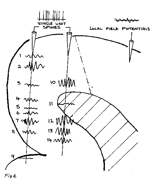
To what level of sophistication can the temporal lobe identify an object? In order to determine that Gross and others have put electrodes into the temporal lobe of primates other than man and determined whether there are neurones which fire maximally when the primate is viewing a specific object. Figure 5 illustrates the main results of this kind of experiment. It turns out that there are in the monkey temporal lobes neurones which respond specifically to the presentation of human faces; indeed these neurones discharge specifically when the faces are presented in profile or face on. The firing rate of neurones is given in the upper part of each panel in the figure as the primate looks at the images given in the lower part of each panel. You can see in this set of images that when the primate is looking directly at the image of another primate face on we get maximal firing but when the image of the head gradually turns around so that it only appears in profile then the firing occurs at a much lower rate. NOW you cannot fool this neurone into firing at a high rate by presenting an image consisting of bits and pieces of a head. If the picture of a brush of the kind used to clean the toilet is presented then the rate of firing of the neurone is much less than that when the image of another primate face on is presented (figure 5). Also if we present an image consisting of the juxtaposition of different elements of the face in a bizarre geometry again the firing rate is not nearly as high as it is when we put those elements together to make up a proper primate face (figure 5). Furthermore, it can be shown that if we take away different elements of the primates face such as the mouth or the eyes then the firing rate will drop (figure 5). Finally this neurone can distinguish the face of a monkey from the human face (figure 5). S6 there are very specific neurones here in the temporal lobe which are specific in the sense that they will fire vigorously only when a particular kind of object, in this case a particular face, is presented. The problem though that we now face is that we might have in temporal lobe neurones for identifying faces but the experience of whether this face is moving across our visual field is not in the temporal lobe but in parietal cortex. As I indicated in figure 4 the problem now is how do we have an holistic experience in our consciousness when the face is moving past us. We have obtained very recently some insight concerning this problem from the work primarily of Wolf Singer working at a Max Planck Institute for Brain Research in Frankfürt. Singer and his colleagues have shown that neurones firing in one part of the brain, concerned for example with the movement of an object such as in parietal cortex, and neurones firing in a different part of the brain, such as in temporal cortex concerned with the fact that the thing that is moving is a face, are both firing at a rate modulated at a frequency of about 40 to 50Hz. Furthermore they fire in phase together not asynchronously (figure 6). There is then a binding together of the firing pattern of those neurones which are activated by the same visual object even though the neurones are located in different parts of the cortex. This discovery is causing a lot of excitement in neurophysiology because it is only those neurones which are firing in phase with this 40 to 50Hz frequency that are giving us the experience of attending to a different part of our visual field. The rest of the visual scene projects from our retina onto the visual cortex at the back of our skulls. The neurones in cur brains subserving those parts of the image on our cortices that is not being attended to are not firing with a modulation of 40 to 50Hz and are not firing in phase.
To some extent it can be claimed that this is all very interesting but that the problem has merely been pushed back one step. You consciousness of phenomena around you might be associated with sets of particular neurones in your brain firing in phase at 40 to 50Hz but how is it that neurones firing off in this pattern give rise to you attending or experiencing a particular facet of your visual world? Only a particular facet of your visual world is entering consciousness. What is there particular about the neurones firing at 40 to 50Hz and in phase which allows you to experience at the conscious level those events which are coded for by these particular neurones? A senior colleague of mine recently commented that this is as far as neurophysiology is going to take US. All that science will illuminate in relationship to your consciousness of the world around you may be that sets of neurones subserving certain specific kinds of functions fire in a certain way. The question of what it is that links consciousness to these sets of neurones is not one which science can answer. If there is any interference with normal brain function at all, for example through disease or injury to the inferior temporal lobe, or through inappropriate development, then we are not capable of consciousness in those areas of experience normally subserved by the injured neurones. (责任编辑:泉水) |
Understanding the Brain in the 21st(2)
时间:2006-03-09 15:48来源:royalsoc.org 作者:bioguider 点击:
1961次
顶一下
(3)
100%
踩一下
(0)
0%
------分隔线----------------------------
- 发表评论
-
- 最新评论 进入详细评论页>>
- 特别推荐
-

- 推荐内容
-