 |
 |
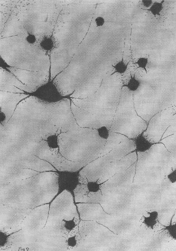
| Figure 9 [Click on image to enlarge] The neurones shown are found in the retina and are called retinal ganglion cells. They connect the retina in the eye to the visual centres of the brain. Many of these neurones degenerate during normal development as the retina is making appropriate connections with the visual centres of brain. Present evidence suggest that this naturally occurring cell death occurs as a result of the neurones failing to obtain a growth factor necessary for their survival. This growth factor is synthesized in the visual centres of the brain where the terminals of retinal ganglion cells obtain the factor and transport it along their axons back to the ganglion cell bodies in the retina. There it is utilized to maintain the integrity of the neurone. |
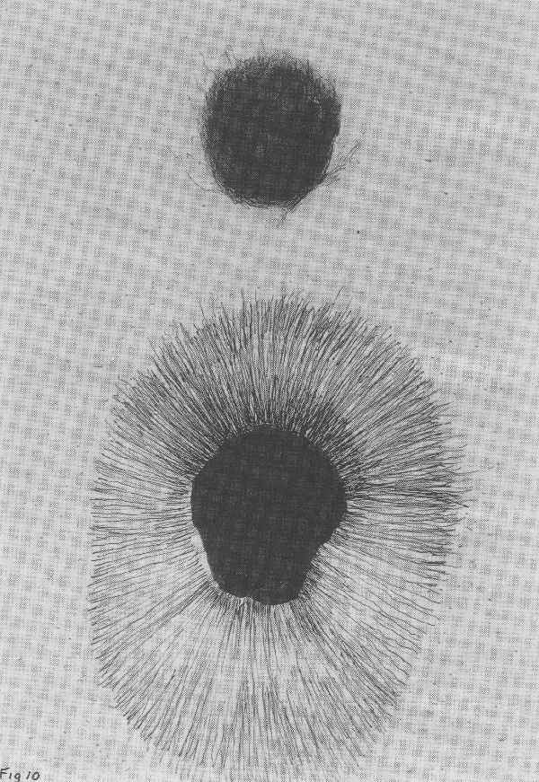
Figure 10 [Click on image to enlarge] Neuronal growth factors for the survival of neurones were first discovered in the peripheral nervous system that controls such organs as blood vessels; these are called sympathetic neurones. Rita Levi-Montalcini and Cohen purified this sympathetic nerve growth factor (NGF) down to a single type of molecule. This figure shows a clump of sympathetic neurone cell bodies in a culture dish in the absence of NGF (upper figure) compared with a clump in the presence of NGF (lower figure). Note that hundreds of axon processes emerge from the clump of neurones in the presence of NGF indicating that these neurones are alive and growing. |
[NextPage]
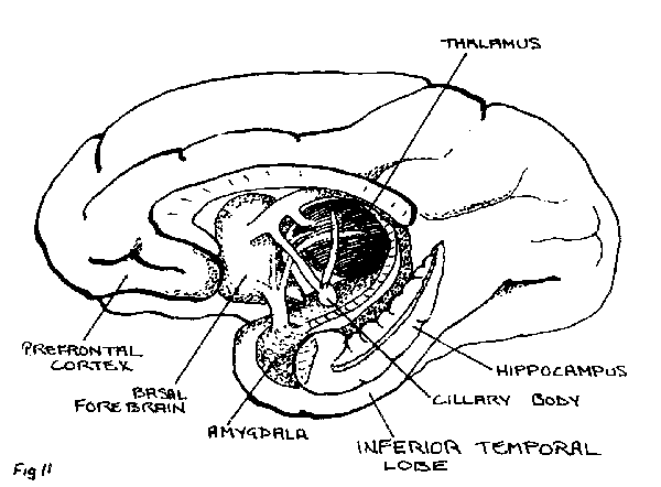
The question then arises as to whether neurones lying deeper in the brain such as those belonging to the temporal lobe and to the hippocampus also have growth factors which will allow for their survival. The neurotrophic factor which keeps retinal ganglion cells alive is not the growth factor that Levi-Montalcini discovered in smooth and cardiac muscle that keeps autonomic neurones alive. The question then is to what extent can we reconstitute an injured temporal lobe or hippocampus by adding in a neurotrophic factor. The hippocampus, a relatively old and primitive type of cortex, abuts onto the temporal lobe (see figure 11). Despite its relative simplicity, the hippocampus is crucially involved in the formation of memories. If a transverse section is cut through this region of the brain many different classes of neurones can be identified (see figures 12 and 13). Some of these are the first neurones to degenerate anywhere in your brain if you have Alzheimer's Disease, particularly those neurones which project into the hippocampus from the part of brain called the septum. Surprisingly, these septal neurones are kept alive by Levi-Montalcini's growth factor, first found to keep autonomic neurones alive in the peripheral nervous system. So if a neurotrophic factor is discovered that saves a certain class of neurones in the periphery it may well work on a class of neurones in the brain.
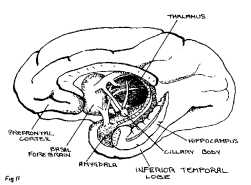 |
| Figure 11 [Click on image to enlarge] The limbic or "border" system of the brain comprises primarily the inferior temporal lobe, hippocampus and amygdala. The hippocampus and amygdala lie on the medial surface of the brain rather than on its lateral aspect. Not only is the system most frequently involved in epileptic fits but it is also one of the first parts of the brain to degenerate in Alzheimer's disease. |
 |
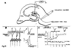 |
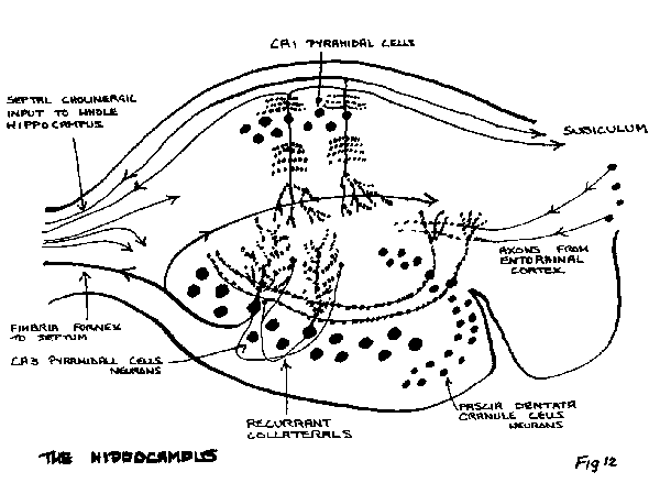
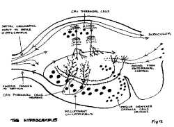
Figure 12 [Click on image to enlarge] A section through the hippocampus, isolated from the rest of the brain. The diagram also shows the layout of the principal neurononal types (indicated as spheres with branching trees of processes called dendrites) together with their interconnections (indicated by the long thin processes, called axons, with arrows attached). The specific input, bringing information about all sensations from the cortex (such as sight, sound or smell), come from the axons arising in entorhinal cortex which synapse on the dendritic processes of granule cell neurones in the so called fascia dentata. These neurones themselves relay this transformed information to the pyramidal neurones in the CA3 region via their axons that form synapses on the CA3 neurones. These neurones are known to code for memories; they connect with each other through processes called recurrent collaterals (only two of which are shown). It is the excitability of these neurones, in part conferred upon them by these recurrent collaterals, that makes this region of the brain the most likely to trigger an epileptic seizure.
The CA3 neurones in turn project to the CAl pyramidal neurones on which they synapse. These neurones also code for memories and their axons project to the region called the subiculum in the old paleocortex and from there to the neocortex. Thus the trisynaptic pathway is from entorhinal cortex to fascia dentata to CA3 pyramids to CA1 pyramids back to the cortex. There is another, less specific input (on the left) necessary for the normal functioning of the hippocampus and that comes from the subcortical structure called the septum. These axons synapse on all neurones in the hippocampus. They are the first neurones to degenerate in the brain of people suffering from Alzheimer's disease. This leads to the loss of memory formation that characterized this disease. |
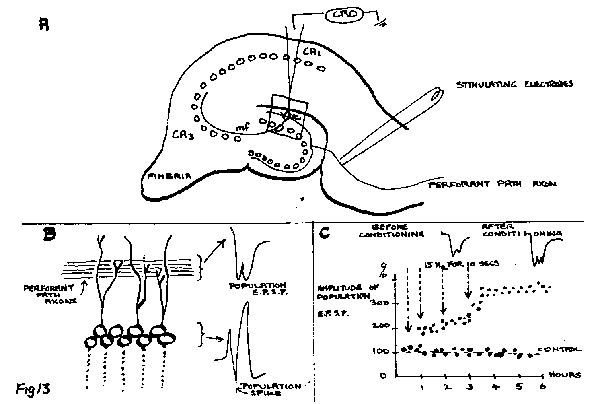
Figure 13. [Click on image to enlarge] The functional circuit between different types of neurones, shown in figure 12, the synaptic connections within this circuit have the very special property of remembering over very long periods of time if they have been subjected to impulse traffic. 'W' shows a transverse section through the hippocampus, like that shown in figure 12, except that stimulating electrodes have been placed on the nerves from the entorhinal cortex (called the perforant pathway axons) and a recording electrode in the dendritic layer of the granule cells, where the perforant pathway axons synapse. "B" shows an enlargement of the boxed area in "A", with sample electrical recordings of the compound population spike occurring because many granule cells fire in synchrony due to stimulating the perforant path axons; the population excitatory postsynaptic potential (epsp) is recorded from the synaptic regions between the perforant path axons and the granule cells an stimulation and gives a measure of the efficacy of synaptic transmission. "C" shows the results of stimulating the perforant pathway at a frequency of 15Hz for 10 seconds at the four times indicated in the graph of the relative amplitude of the population epsp against time; if the perforant nerves are stimulated every few minutes with a single impulse before, during and after the 15HZ stimulating periods then the amplitude of the population epsp at each 6 minute period is shown to grow over 3 hours until it settles down to a size 300% that of the control (in which no stimulation occurred at 15Hz); two examples of this long-term potentiation of the population epsp are shown one before and one after the 15HZ conditioning period, This enhanced efficacy of transmission through the synapses of over 300% is shown to last for 3 hours after the I5Hz stimulation but may continue for days or months. The mechanisms responsible for this potentiation are required to retain a memory. |
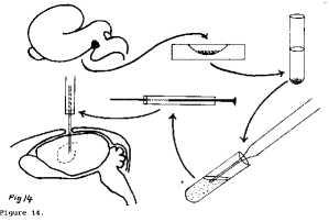 |
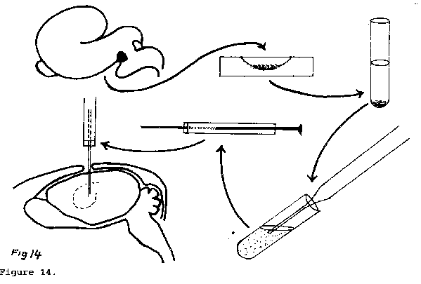
| Figure 14. [Click on image to enlarge] Procedure for transplanting embryonic septal neurones from the hippocampal region of a foetus to the hippocampus of a mature animal with a degenerating septum. The septal region (black area) is first removed from the foetal brain and placed in a culture dish with enzymes that loosen the tissue into separate neurones. The partly separated neurones are then placed in a test tube and the isolation process taken further by rapidly shaking the neurones up and down in a pipette in the test tube. The completely dissociated neurones are next taken up into a syringe. The syringe needle is then located with great accuracy in the appropriate part of the hippocampus and the dissociated septal neurones injected into this area of the brain. |
As already mentioned, rather than introducing growth factors into the brain in the hope of saving a certain class of neurones that are degenerating because of a neurological disease, it is possible to introduce embryonic neurones of the same class that have been destroyed by the disease (figure 14). These can be obtained from a set of neurones that have been genetically manipulated to be of a similar kind to those that have degenerated. If you introduce these neurones into the brain (see figure 14), they reconstitute the normal circuitry, making the correct functional connections. In this way they restore the normal function, or at lease almost the normal function, of that part of the brain which had degenerated.
(责任编辑:泉水) |