
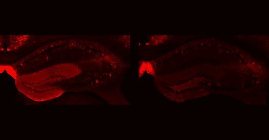
前路分歧:一项发表于《Nature》的最新研究表明,缺乏TSC2基因的人类大脑类器官会产生过度活跃的星形胶质细胞,其特征与结节性硬化症复合物(TSC)患者脑内结节中发现的细胞高度相似。
这一研究结果强有力地证实了一个观点:胶质细胞的病理变化是由蛋白质表达和活性的改变——具体而言是高水平的mTORC1信号传导——所驱动的,而不是由癫痫发作本身继发引起的。研究人员在论文中写道:“这些结果暗示胶质细胞功能障碍是TSC发病机制的驱动因素,并强调反应性星形胶质细胞有望成为TSC相关神经病理学潜在的治疗靶点。”
本周精选自闭症与神经发育研究进展:
除了上述突破性成果外,近期学术界在神经发育与精神疾病领域还涌现出多项重要预印本与期刊论文:
• “Postnatal maturation of putamen microstructure accompanies topographic white matter connectivity and altered circuits in autism” 详见 bioRxiv。
• “A retinoic acid autoregulatory loop governing prefrontal-motor arealization” 详见 Nature。
• “Developmental cerebellar pathology in mouse models of SCN2A premature termination codon variants” 详见 bioRxiv。
• “Maternal-fetal immune conflict contributes to male-specific impairments in a mouse model of neurodevelopmental disorders” 详见 Science Advances(相关解读可参考:“Post-infection immune conflict alters fetal development in some male mice”)。
• “Parvalbumin interneurons and dentate gyrus homeostatic dysregulation shape epileptogenesis in Angelman syndrome model mice” 详见 bioRxiv。
1. Organoids missing the TSC2 gene form rogue astrocytes. DOI: 10.1038/s41586-026-11054-w
2. Postnatal maturation of putamen microstructure accompanies topographic white matter connectivity and altered circuits in autism. DOI: 10.64898/2026.09.09.750447
3. A retinoic acid autoregulatory loop governing prefrontal-motor arealization. DOI: 10.1038/s41586-026-11014-4
4. Developmental cerebellar pathology in mouse models of SCN2A premature termination codon variants. DOI: 10.64898/2026.09.13.751275
5. Maternal-fetal immune conflict contributes to male-specific impairments in a mouse model of neurodevelopmental disorders. DOI: 10.1126/sciadv.aeg0779
6. Parvalbumin interneurons and dentate gyrus homeostatic dysregulation shape epileptogenesis in Angelman syndrome model mice. DOI: 10.64898/2026.09.17.752419